Applications - LifeScience Gallery
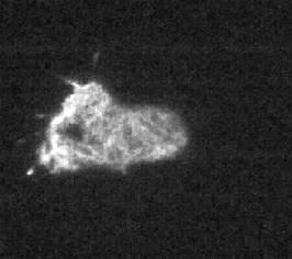
TIRF microscopy image taken with the SensiCam VGA and Image J software.
Courtesy of Imperial College London, England.
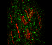
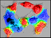
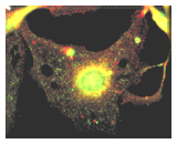
Green dots are terminals from glutamate neurons. The red cells are the neurons
that regulate LH and FSH secretion from the pituitary and trigger ovulation.
Images captured using the SensiCamQE High Performance Digital CCD Camera. Click
here to view larger version movie clip (3.2MB). Images courtesy of the University
of Maryland, Gloria E. Hoffman, PH.D., Department of Anatomy and Neurobiology.
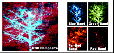
Images captured using the DiCam-PRO Intensified Digital CCD Camera. Laser Induced
Fluorescence Imaging Classification algorithms and ratio imagery are useful
in isolating fluorescence emissions from plant components of scientific interest.
Images courtesy of SSAI USDA ARS Hydrology and Remote Sensing Lab Lawrence A.
Corp-Sensor System Specialist - Beltsville MD
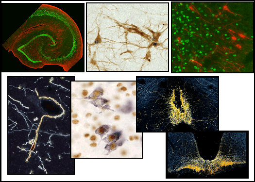
Images captured using the SensiCamQE High Performance Digital CCD Camera. Click here for more details. Images courtesy of the University of Maryland, Gloria E. Hoffman, PH.D., Department of Anatomy and Neurobiology.
HELA cells stained with the Calcium indicator Fura-2. Courtesy of T.I.L.L Photonics GmbH, Germany
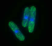
Cell Imaging: A deconvolved and 2D projected image of fission yeast stained for microtubules.
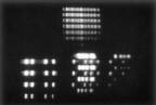 Chemiluminescence
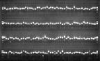 Northern
Blot
Northern
Blot
 Multiprobe
Fluorescence Imaging
Multiprobe
Fluorescence Imaging
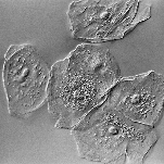 Cheek Cells
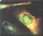
 Multiprobe
Fluorescence Image
Multiprobe
Fluorescence Image
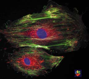
 Fibroblasts
Fluorescence Imaging
Fibroblasts
Fluorescence Imaging
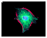
 Melanocytes
Fluorescence Imaging
Melanocytes
Fluorescence Imaging